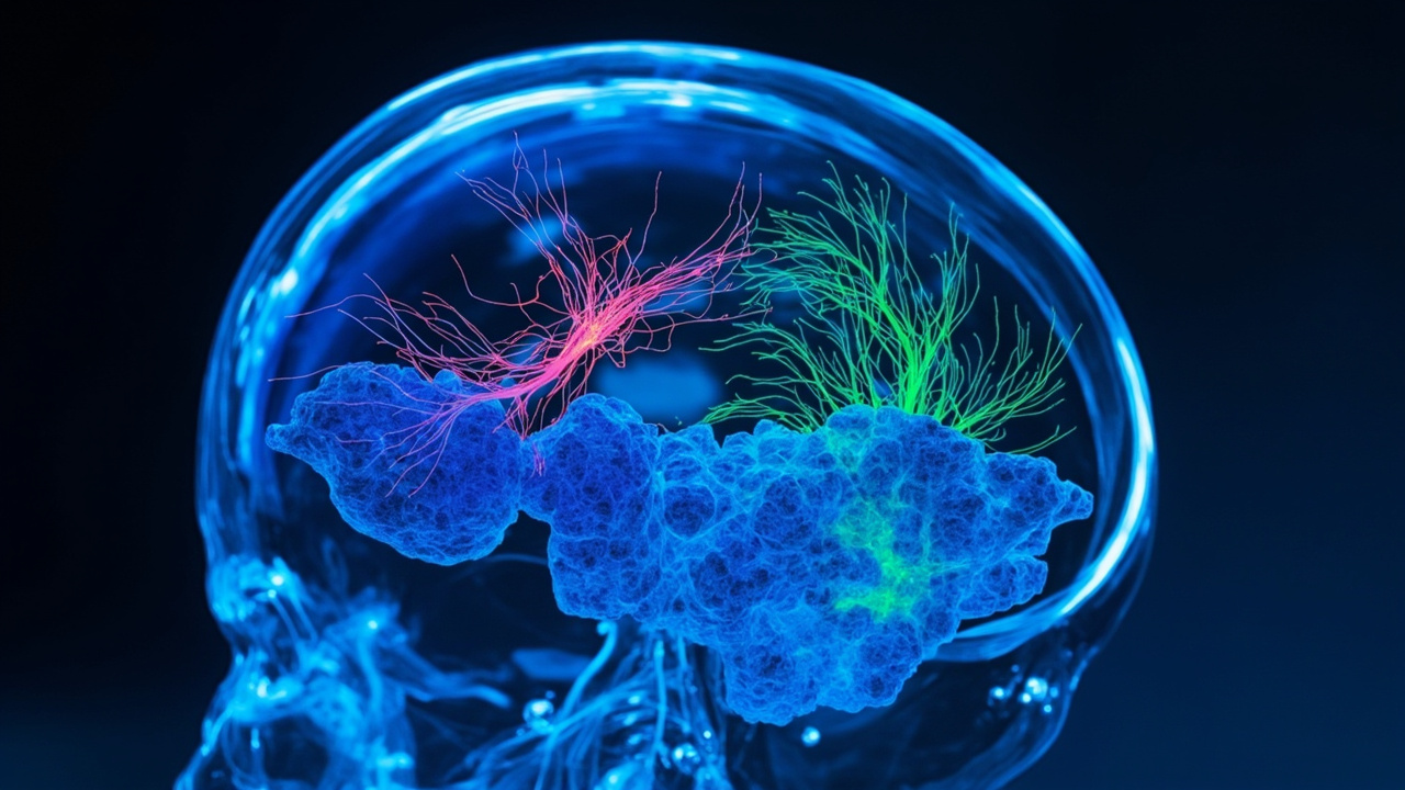
TL;DR: On September 16, 2026, Stanford scientists reported in Nature that lab-grown human cortical organoids transplanted into mice born without most of their cortex grew to fill the empty space and formed functional connections.
Stanford University neuroscientist Sergiu Pașca led a team that created so-called xenocortical mice to study human brain development in a living nervous system, according to Reuters in its report titled "Scientists transplant lab-grown human brain tissue into mice."
The study was published on September 16 in the journal Nature by Konstantin Kaganovsky, Kevin W. Kelley, Tilo Gschwind, Paul M. Harary and colleagues, with Pașca as senior author. The paper is titled "Developmental xenocortication using human-derived organoids in mice."
Why living human brain tissue is hard to study
Living human brain tissue is essentially inaccessible for research for ethical reasons, the researchers said, which limits study of human development, circuit function and disease mechanisms.
Human stem-cell-derived models are beginning to address that gap, according to the Nature paper abstract. Three-dimensional cortical organoids grown from human induced pluripotent stem cells can recapitulate aspects of neurogenesis and gliogenesis of the cerebral cortex in laboratory dishes.
The idea of growing miniature brain structures in a laboratory dish emerged in the early 2010s, when researchers in Austria showed that stem cells could self-organize into three-dimensional tissue resembling aspects of the developing human brain, according to Science News in its report titled "These mice have human (nerve cells) on the brain."
But those clusters, called organoids, lacked the connections, blood supply, sensory inputs and motor outputs of a living brain. As Pașca explained in that coverage, the goal has been to make aspects of human brain development and function accessible for investigation so therapeutics can be developed.
How the team made room for human tissue
In 2022, Pașca and colleagues developed a way to transplant human organoids into newborn rats, eventually getting human cells to make up about one-third of one side of the rat's cortex. But rat neurons mature faster than human counterparts, limiting how much the human tissue could develop in that rodent model.
To overcome space constraints and competition with host circuits, the Stanford group used a genetic strategy to deplete glutamatergic neurons from the mouse neocortex and hippocampus. The resulting animals were termed apallial mice.
The bioengineered mice lacked 98% of the cortical and hippocampal tissue found in ordinary mice, according to Reuters. Those two structures are involved in movement, learning and memory and together account for about half of the brain's total volume.
Although the resulting animals showed some behavioral defects, they were described as surprisingly functional. Anatomical studies, magnetic resonance imaging and whole-brain single-nucleus RNA-sequencing confirmed that apallial mice lack dorsal and medial pallium derivatives, including neocortex and hippocampus.
How the human organoids were grown
The researchers fashioned miniaturized, three-dimensional organoids designed to reproduce key cell types and developmental features of the human cerebral cortex, the brain's outermost layer governing cognition, language, attention and decision-making.
They did so by reprogramming skin or blood cells to become stem cells capable of transforming into almost any cell type in the body, then guiding those cells to form self-organizing clusters of cortical tissue.
According to Scientific American in its report titled "Human brain organoid transplants mesh with mouse brains, forming active neural circuits," the team placed four organoids into each mouse's empty intracranial space, with each organoid containing about 100,000 neurons at the time of transplant.
The transplant was performed shortly after birth, placing human stem-cell-derived cortical organoids into the cortical cavity. The space normally occupied by the mouse cortex gave the human tissue room to grow extensively.
What grew inside the xenocortical mice
Transplantation into apallial mice, termed xenocortication, resulted in growth of a large volume of human-derived neural tissue, according to the Nature abstract. The researchers called the grafted animals xenocortical, or XCX, mice, referring to the foreign transplanted human cortical tissue.
The transplantation successfully took hold in 25 of 29 attempts, according to Science News. Over the next three months, the human tissue expanded nearly fivefold in volume and ultimately filled more than 90 percent of the cortex.
Within a few months, those starting populations had divided into roughly four million neurons, according to Scientific American. The cells sent axons — fibers that convey electrical signals — throughout the mouse nervous system all the way to the spinal column.
The human grafts generated a broad diversity of cortical cell types and established functional connections throughout the mouse nervous system. Those types included layer 5 extratelencephalic projection neurons and a rare type called von Economo neurons, considered vulnerable in some forms of dementia and thought to be important for social cognition in primates, elephants and dolphins.
These cells have never been documented in organoids confined to a petri dish, suggesting that the mouse-brain environment enables human neurons to develop more fully. The tissue lacked some features of a mature cortex, including its characteristic layers, yet still wired extensively into the mouse nervous system.
Electrical activity resembled developing circuits
Human cortical neurons integrated with the mouse nervous system, and in vivo cortical graft-wide calcium imaging and electrophysiological analyses revealed patterns of organized activity resembling developing circuits, according to the Nature abstract.
Brain imaging showed patterns of coordinated activity across the human graft. Pașca said this tells researchers that the cells are active and communicating with each other, according to Scientific American.
The researchers emphasized terminology. Pașca said descriptions such as humanized mice, mice with human brains or mini-brains are not accurate representations of what was created. The animals retain a mouse nervous system but contain a larger volume of human cortical tissue that develops, integrates and forms connections within it.
Pașca also said cortical organoids give researchers an experimental window into human brain development and disease. They are not miniature brains and do not reproduce the full complexity of the human brain, but they allow study of human neural cell types and developmental processes that would otherwise be extremely difficult to access.
Behavior was broadly preserved with selective differences
Behavioral analyses of apallial and xenocortical mice revealed broadly preserved locomotion alongside selective differences in limb coordination and altered organization of spontaneous behavior, according to the Nature abstract.
Unsupervised machine learning applied to spontaneous mouse behavior using motion sequencing, known as MoSeq, revealed distinct behavioral repertoires in control and apallial mice, with the XCX group positioned intermediate between, yet distinct from, both groups.
Targeted tasks demonstrated minimal gross impairments. However, apallial mice showed deficits on tasks requiring neocortex and hippocampus, including fine motor coordination in the CatWalk test and working memory in the Y maze, a pattern that was only partially observed in XCX mice.
In other words, on tests of fine motor control and memory, the grafted mice's performances fell somewhere between that of mice with intact brains and those lacking a cortex. One outside researcher not involved in the work noted that a mouse without a cortex is relatively sick, and after transplant it behaves more like a normal mouse.
Oxygen deprivation modeled injury linked to cerebral palsy
As a first application, the researchers exposed the mice to oxygen deprivation to model injury to developing human cortical cells.
The developing human brain can be highly vulnerable to oxygen deprivation. When it occurs during pregnancy or around birth, it can have major neurological consequences including contributing to cerebral palsy and increasing the risk of epilepsy or autism, Pașca said. Mice can be resilient to levels of oxygen deprivation that injure human neural tissue.
After oxygen deprivation, the human tissue showed cellular signs of injury, and the mice developed problems with gait and limb coordination — features of the disability in people as well, according to Science News. Moreover, hypoxia induced a cellular response along with deficits in the CatWalk procedure in XCX mice, underscoring the potential of the platform to facilitate disease modeling and therapeutic discovery using large human-derived grafts and behavioral outputs.
Pașca said this approach could be used down the line to test many therapeutics being considered for cerebral palsy. He plans to study frontotemporal dementia and genetic forms of autism with the mice, using organoids made from patient-derived cells affected by those conditions.
What this could mean for autism, epilepsy and schizophrenia
The researchers said the human tissue replicated key features of brain development including formation of functional neural networks after transplantation. Pașca said the work gives researchers a way to study human neural tissue across levels, from genes and individual cell types to circuits and functional consequences in an animal.
The new methodology should speed research into the underlying biological causes of schizophrenia, epilepsy, profound autism and cerebral palsy, Pașca said. Neuroscientists will be able to learn more about causes and mechanisms of neurodevelopmental and pregnancy-incurred disorders and to test possible interventions.
This approach could be especially useful for studying disorders that begin during brain development such as autism, epilepsy, schizophrenia and cerebral palsy, as well as environmental injuries such as oxygen deprivation around birth, according to the researchers.
Pașca said the team built a system so that it captures aspects of disease that would be difficult to capture with other systems. Outside neuroscientist Simon Hippenmeyer, who was not involved, called the result absolutely transformative and said it promises future applications for studying development and modeling neurodevelopmental diseases.
Outside scientists weigh promise and ethics
The technique is certainly bold, said H. Isaac Chen, a neurosurgeon at the University of Pennsylvania who studies brain organoids but was not involved in the research, according to Science News. The extent of humanization raises questions about ethical boundaries, Chen noted, but Pașca has gone to unusual lengths to grapple with those concerns.
Pașca consulted an outside panel of experts led by Insoo Hyun, a bioethicist at the National University of Singapore Yong Loo Lin School of Medicine. Hyun acknowledged the model raises legitimate questions about where to draw the ethical line, but said there is little evidence these mice crossed it.
What matters, Hyun said, is not how much human tissue an animal contains, but what that tissue enables it to do. From a secular ethical point of view, he said, it boils down to cognitive capacity — and so far these mice do not seem to have gained obvious extra brainpower.
Geneticist Alysson Muotri, who was not involved, called the work a tour de force in terms of technology, according to Scientific American, but argued it is hard to disentangle human neuron activity from rodent neuron activity, which could complicate efforts to probe human brain function.
Sources referenced
- Scientists transplant lab-grown human brain tissue into mice | Reuters (https://www.reuters.com/business/healthcare-pharmaceuticals/scientists-transplant-lab-grown-human-brain-tissue-into-mice-2026-09-16/)
- These mice have human (nerve cells) on the brain - Science News (https://www.sciencenews.org/article/mice-brain-human-organoid-cells)